3D X-ray microscopy (XRM)

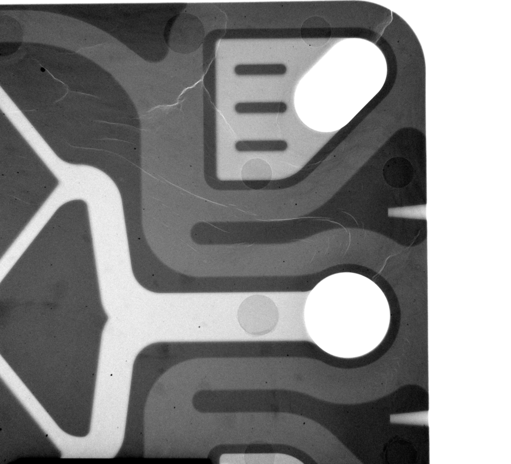
The ZBT is equipped with the ZEISS Xradia 620 Versa 3D X-ray microscope, a high-resolution system for non-destructive material and component analysis. 3D X-ray microscopy provides detailed insights into the interior of samples and reveals structures that are only partially accessible using conventional two-dimensional or destructive methods.

At the ZBT, the system is used primarily to investigate materials and components for hydrogen technologies. These include fuel cell components, porous transport layers, membrane electrode assemblies, injection-moulded parts, and additively manufactured structures. The method combines high-resolution imaging with quantitative structural, defect, and damage analysis.
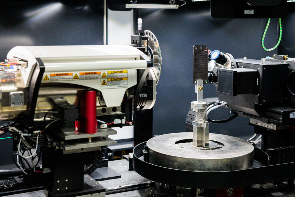
3D X-ray microscope at the ZBT - The Hydrogen and Fuel Cell Center for the investigation of hydrogen technologies

Applications of 3D X-ray microscopy

XRM measurements allow material structures to be captured, visualised, and quantitatively analysed in 3D without causing damage. The method supports both scientific research and industrial development and quality assurance processes.

Typical areas of application at the ZBT include:

  • Structural Analysis of Porous Materials
  • Investigation of Fuel Cell and Electrolyser Components
  • Quality Control of Injection-Moulded or Additively Manufactured Components
  • Analysis of Defects, Cracks, and Inhomogeneities
  • Study of Degradation, Frost Damage, and Aging Effects
  • Data Set for Structure-Resolved Simulations

What can be analysed using XRM?

Realistic 3D Structures for Simulation and Modeling

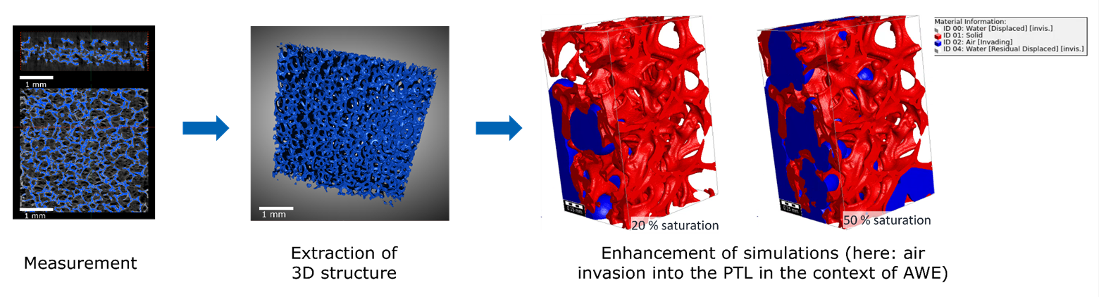
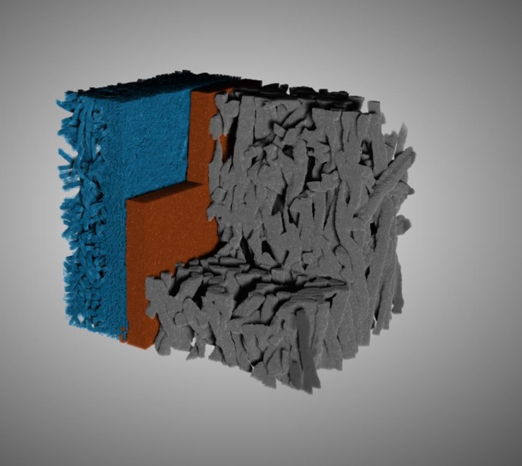
Porous structures influence mass transport, water distribution, gas transport, and performance in electrochemical systems. Using 3D X-ray microscopy, real-world pore geometries can be measured and utilised for structure-resolved simulations.

This allows idealised or highly simplified models to be replaced by measured 3D structures. As a result, transport pathways, local saturation, bottlenecks, and performance-limiting areas can be analysed more accurately. At the ZBT, this approach has been used, among other things, for complex porous components and interfaces in fuel cell and electrolyser contexts.

Quality Control of Injection Moulding and Manufacturing Processes

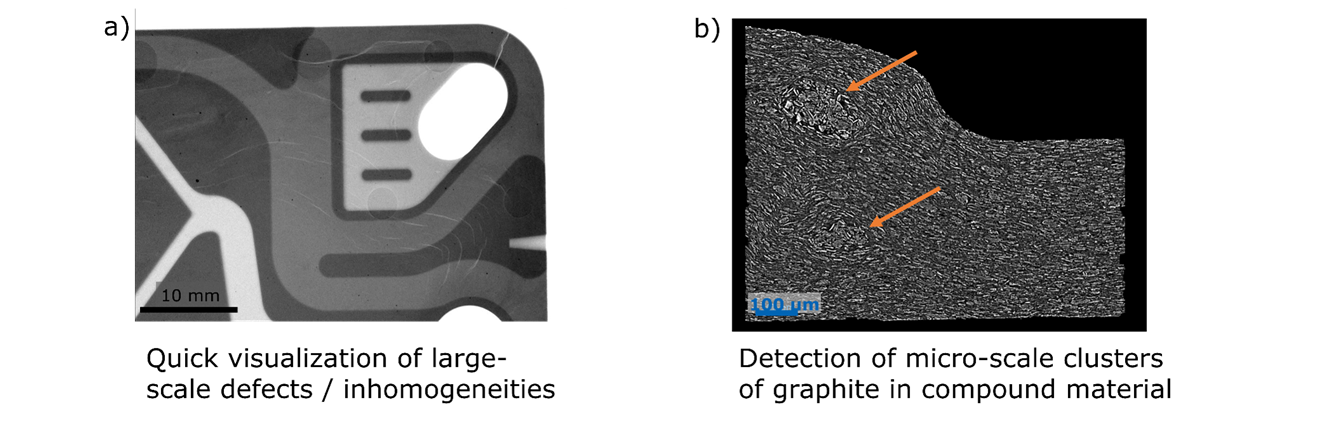
XRM measurements enable the nondestructive inspection of technical components across various length scales. In injection-moulded components, they can reveal internal inhomogeneities, defects, filler distributions, or particle orientations.

This information can be utilised to optimise manufacturing processes in specific ways, such as with regard to temperature, pressure, speed, or material composition. At the same time, the analysis of the microstructure helps provide a better understanding of material properties such as thermal conductivity or mechanical stability.

Analysis of Degradation and Frost Damage in Fuel Cells

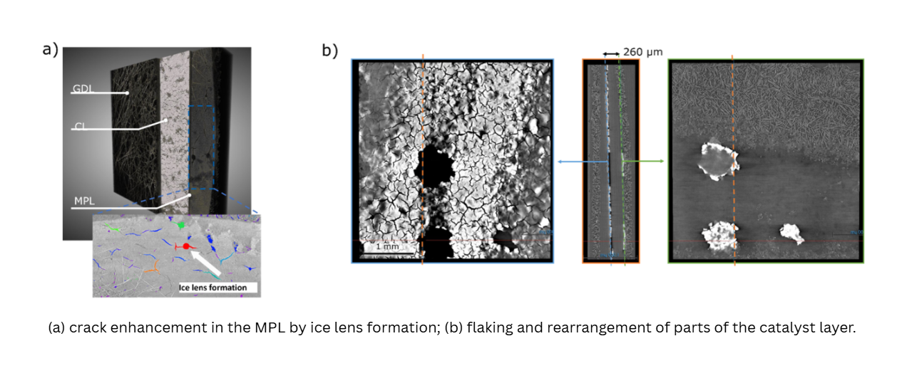
Fuel cell components are subjected to demanding conditions during operation. Cold start conditions and freeze-thaw cycles, in particular, can alter the material structure of membrane electrode assemblies and adjacent layers.

3D X-ray microscopy makes it possible to examine such changes within the multilayer system non-destructively. For example, it reveals cracking, delamination, structural rearrangements, or damage caused by moisture or ice. This allows for a better understanding of aging mechanisms and enables the targeted further development of materials, components, and operating strategies.

What can be analysed using XRM?

3D X-ray microscopy makes it possible to visualise, measure, and evaluate internal structures and material properties. Depending on the sample and the research question, qualitative visualisations, quantitative structural parameters, or data-driven analyses can be generated.

Among other things, the following can be examined:

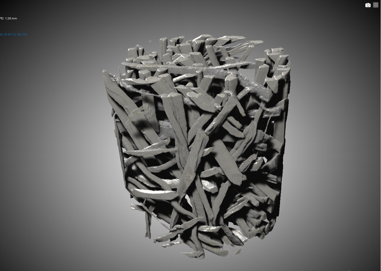
  • Pore Structures, Porosity, and Pore Size Distributions
  • Cracks, delaminations, and internal defects
  • Layer Structure and Structural Changes in Multilayer Systems
  • Particle, fiber, or filler orientations
  • Water Distribution and Transport Pathways in Appropriate Sample Setups
  • Internal geometries of technical components
  • Damage caused by aging, frost, and moisture
  • additively manufactured structures, nozzles, composite materials, and engineering samples with complex internal geometries

Technical Specifications

For 3D X-ray microscopy, the ZBT utilises the ZEISS Xradia 620 Versa. The system is suitable for high-resolution 3D analyses of engineering materials and components and can be used for a variety of sample sizes and research questions.

  • Maximum resolution: up to 0.5 µm
  • Power: 25 W
  • Voltage range: 30–160 kV
  • Available Objectives: 0.4x, 4x, 20x, 40x
  • Maximum sample weight: 25 kg
  • In-situ measurements are possible

Measuring principle

  • Radiography: X-rays transmitted through the sample are converted into visible light and projected onto a CCD camera.

  • Tomography: X-rays are taken from various angles of rotation to calculate a 3D structure.

  • Software: Data analysis using statistical methods, segmentation, or machine learning algorithms.

3D X-ray microscope

Simon Dondrup
+49 203 7598-2364
Portrait of Simon Dondrup.

3D X-ray microscope

Dr. Viktor Mackert
+49 203 7598-2370
Portrait eines Manns mit Brille, Anzug und Krawatte